

If those reading this and seeing a crisp, clear screen, thank your fovea.
Fovea, Latin for “pit”, is a depression in the back of the eye where visual acuity is sharpest, thanks to a high density of light and color sensing cone cells. Without the fovea, a person couldn’t swipe through an app on a smartphone or drive a car. It is also one of the areas worst affected in age-related macular degeneration (AMD), the leading cause of vision loss in older adults.
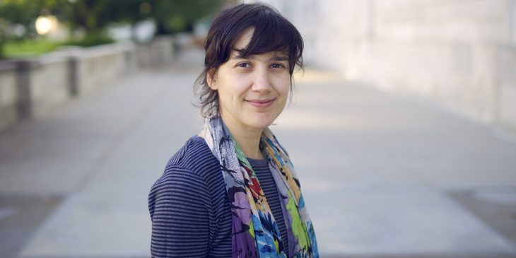
The fovea was described in the early 19th century, so Dr. Susana da Silva, assistant professor of ophthalmology at the University of Pittsburgh School of Medicine, was surprised to learn that no one actually knew much about how it develops in embryos. So, she decided to study it during her postdoctoral research stint at Harvard University.
“Eye doctors care so much about the fovea because it is central to good vision,” said da Silva, who is the newest recruit at the Department of Ophthalmology. “Yet, from a basic biology standpoint, it’s still quite a mystery as to how it develops. I thought it seemed like a very interesting area to explore.”
To study the fovea, da Silva turned to chick embryos, a model system that has been used for centuries to learn about embryonic development. Chickens don’t have a fovea per se, but have a region of high acuity that is similar to the human fovea in that they have a high density of cones and no rod cells, which are responsible for good vision in dim light.
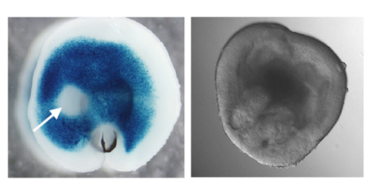
Embryonic chick retina explant (left) showing absence of retinoic acid (blue) in the high acuity area (arrow), and a 50-day old human ‘eye in a dish’ culture derived from induced pluripotent stem cells. (Susana da Silva/Pitt Health Sciences)
Da Silva’s research revealed a surprising new role for retinoic acid, a chemical derivative of vitamin A known to be important in controlling embryonic development. She found that it was actually a lack of retinoic acid that was needed for the high acuity region to form.
“This was the first time that a molecular mechanism responsible for foveal development had been identified,” she said, “and it opens up lots of new avenues for research with implications for treating vision diseases.”
At Pitt, da Silva plans to continue exploring how retinoic acid works in foveal development and is excited about the opportunities for basic and translational research.
“There is a breadth of vision and neuroscience research at Pitt and the environment is really collaborative and supportive. I’m looking forward to some very fruitful collaborations,” da Silva said.
Going beyond chickens, she is also working on a model that is more reflective of how the human eye develops, using stem cells to create a three-dimensional organoid “eye in a dish”. By recreating foveal development in organoids, she hopes to not only understand its biology further, but also test new stem cell-based therapies to restore vision.
“You can count the number of people in the world studying the development of the fovea on one hand, and Susana is one of them.,” said Dr. Jeff Gross, professor of ophthalmology at Pitt’s School of Medicine and director of the Louis J. Fox Center for Vision Restoration, who helped recruit da Silva. “She’s an exceptional scientist and we’re excited to see her build her research program here at Pitt, and for the impact it will have on people’s lives.”
Related Posts:


![]()
![]()
![]()
![]()
![]()
![]()
![]()