

To better understand glaucoma, a leading cause of irreversible blindness worldwide, I and my fellow researchers at the University of Pittsburgh and Harvard School of Medicine have visualized previously unseen elements of the eye. These stunning images reveal new details about the stars of the show: astrocytes, which normally keep nerve cells of the eye healthy. In glaucoma, the role of these star-shaped cells becomes more complicated.
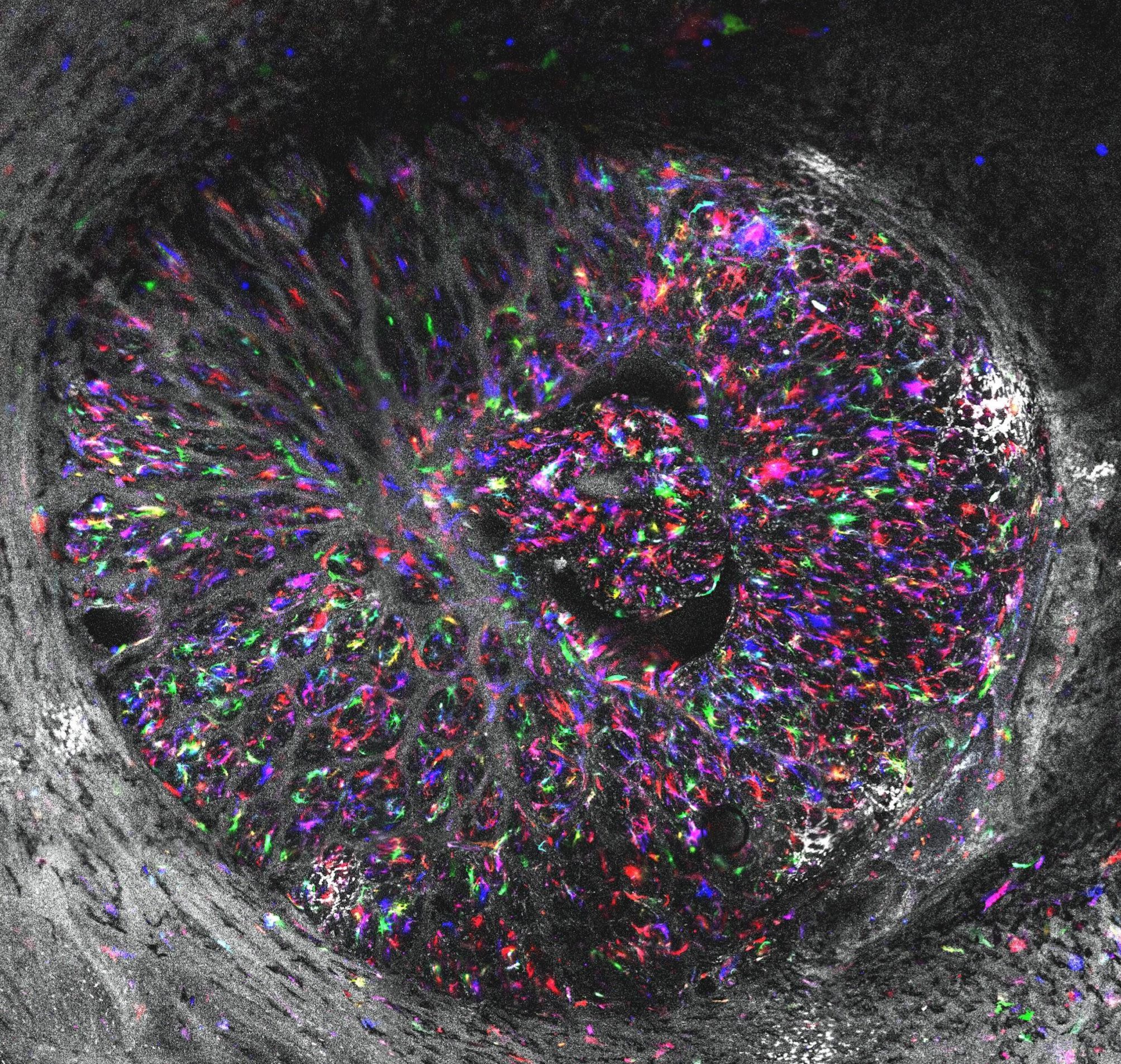
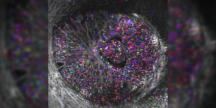
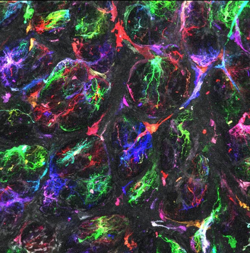
Star-shaped astrocytes across the back of the eye shown in brilliant color using a method called Multicolor DiOlistic labeling.
At the front of the bridge between the eye and the rest of the brain is a region called the lamina cribrosa. This spot is an initial site of the neurodegeneration that causes progressive vision loss in glaucoma. This part of the eye is also densely populated by astrocytes. Lamina cribrosa astrocytes are thought to be early actors in glaucoma, with changes in their shape related to their ability to support neural tissue health. In glaucoma, it is not yet clear whether these astrocytes are heroes, villains, or something in between.
From rodent models, we know that these changes in astrocyte morphology can be detected before any neurodegeneration sets in. But glaucoma research in rodents has serious limitations. For starters, mice and rats do not have a lamina cribrosa as do the eyes of other larger animals and humans. Therefore, our knowledge about astrocyte morphologies in this region is severely limited.
Research in human eyes has often relied on looking at limited parts of astrocytes, such as aspects of their cytoskeleton, the skeleton-like network of proteins that gives cells structured shape. Inferring astrocyte shape based on their cytoskeleton is a lot like imagining what dinosaurs looked like based only on what paleontologists know of their bones.
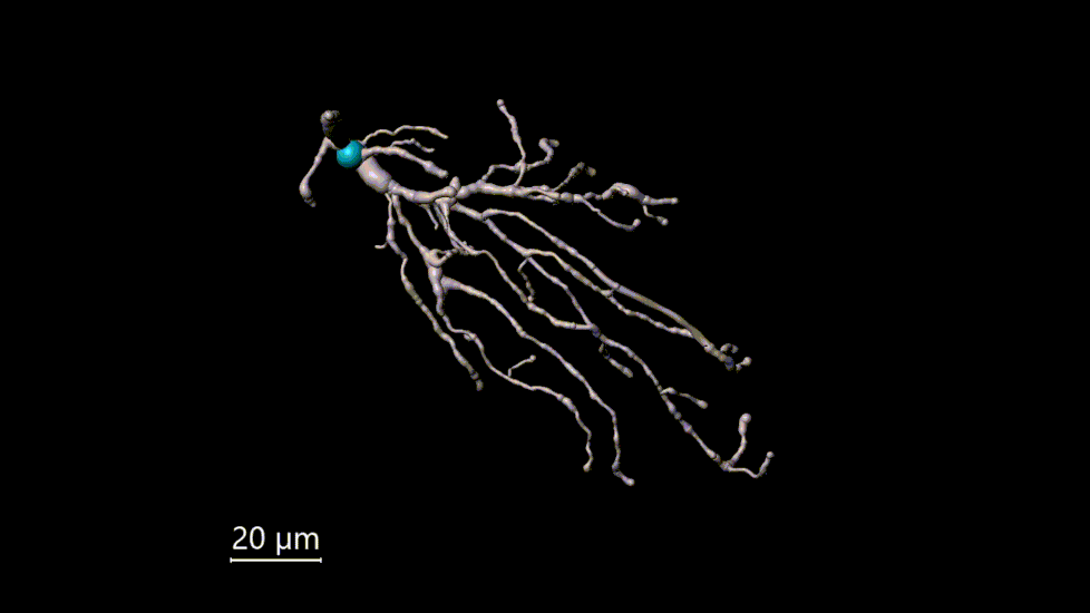
Astrocyte model shown in 3D
In a paper published in late March in Experimental Eye Research, we describe an approach to visualize astrocytes in the eye that does not rely on rodent models or imperfect inferences based on cytoskeleton shape.
Individual astrocytes and details of their morphologies are revealed with 3D microscopy
The approach, called Multicolor DiOlistic labeling, allows us to see astrocytes at unprecedented scale and detail. We used high resolution images of the astrocytes to create an atlas of three-dimensional astrocyte models, enabling automated quantification of astrocyte morphologies.
We aim to use this approach to investigate differences between healthy astrocytes and those in the early stages of glaucoma. This will help us better understand astrocyte-mediated mechanisms of disease and the complicated roles they play.
Susannah Waxman is lead author of this study and a doctoral student in the Laboratory of Ocular Biomechanics at the University of Pittsburgh.
Related Posts:


![]()
![]()
![]()
![]()
![]()
![]()
![]()