In a paper published today in Nature Communications, Dr. Marco Capogrosso, assistant professor in the Department of Neurological Surgery at the University of Pittsburgh School of Medicine, proposed a new technology to improve arm and hand movements in individuals with arm paralysis due to spinal cord injury, stroke or other movement disorders.
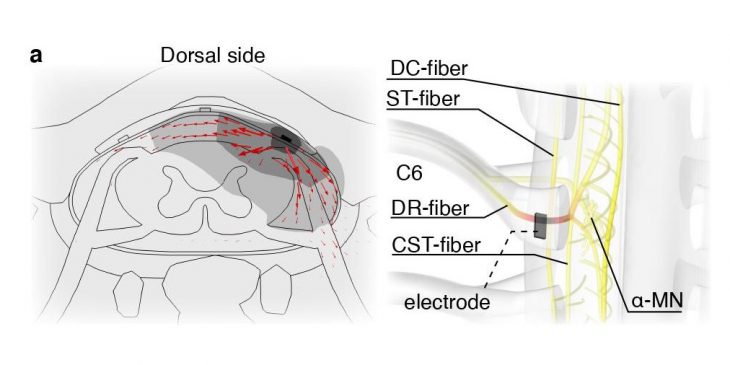
In collaboration with colleagues in Switzerland, Capogrosso and his team combined physics simulations with experiments in macaque monkeys and humans to demonstrate that epidural electrical stimulation of specific regions in the spinal cord can be used to activate arm and hand muscles.
“This work is very important for clinical applications,” said Capogrosso, senior author of the paper, who is also part of the Pitt Rehab Neural Engineering Labs. “To design effective therapies for paralysis, we need to selectively engage hand and arm muscles by providing the surviving neural circuits with appropriate electrical signals. In practice, this means that we need to know where to implant electrodes and how to design them – and our work provides guidance to that.”
Epidural stimulation is a neurotechnology that is used to treat pain caused by damage or injury to the nerves in thousands of patients every year. For this procedure, a surgeon implants a small neurostimulation device consisting of thin electrode wires over the spinal cord’s protective coating. When the device is turned on, it can send electric signals to the damaged nerve fibers and stimulate them, restoring the spinal circuits and dampening the feeling of pain after the injury.
However, modern spinal cord stimulators are not intended to target the cervical neural circuits necessary for arm and hand movement. Therefore, the scientists proposed and tested a modified design of multi-electrode spinal implants that were fitted to monkeys’ spinal cord dimensions.
To determine the optimal placement of electrodes in the cervical spinal cord — where neural circuits that control arm movements are located — researchers used insights from in silico, or computer, experiments and reproduced them in monkeys. Combining computer simulation with animal studies allowed researchers to reduce the number of animals needed to collect meaningful data as part of an effort called the “Three Rs” (Replacement, Reduction, Refinement) that strives to minimize the use of animals in research. And, because the monkey’s cervical spine is anatomically and functionally similar to the human’s, scientists were able to approximate the arm and hand movement in people very closely.
“I think our graduate students Nathan Greiner, Beatrice Barra and the other members of the team did great work in building up from basic science concepts to design this practical technology while minimizing the number of animals used in this research,” added Capogrosso.
However, there is still significant work to do to optimize this technology for use in people.
Capogrosso moved to Pittsburgh in January 2020 and joined the Department of Neurological Surgery to use the impressive resources of UPMC to bring this technology to patients as soon as possible. Thanks to collaborations with neurosurgeons such as Dr. Peter Gerszten, Capogrosso hopes to start testing the implant in patients soon.
Using the tools developed by other researchers, such as Dr. Fang-Cheng Yeh at the Department of Neurological Surgery and Dr. Elvira Pirondini at the Department of Physical Medicine and Rehabilitation, they will use precise MRI imaging to target electrode implantation in humans and reproduce the results obtained in monkeys.
“Hopefully, we’ll see the first results in humans in 2021,” Capogrosso said.


